Home

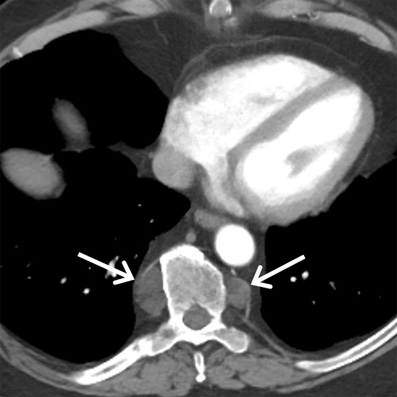
paraspinal soft tissues are normal

Imaging Soft Tissues of the Neck | Radiology Key

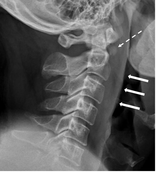
Normal prevertebral soft tissues. Normal lateral radiographs (A, B).... |  Download Scientific Diagram
Normal prevertebral soft tissues. Normal lateral radiographs (A, B).... | Download Scientific Diagram

Neck pain with prevertebral soft tissue thickening - Annals Singapore

Understanding a mass in the paraspinal region: an anatomical approach |  Insights into Imaging | Full Text
Understanding a mass in the paraspinal region: an anatomical approach | Insights into Imaging | Full Text

Imaging of the Posterior/Paravertebral Mediastinum | Radiology Key

Differentiating Normal from Abnormal Inferior Thoracic Paravertebral Soft  Tissues on Chest Radiography in Children | AJR
Differentiating Normal from Abnormal Inferior Thoracic Paravertebral Soft Tissues on Chest Radiography in Children | AJR

A) 59-year-old woman, paravertebral soft tissue type. (A, B and C)... |  Download Scientific Diagram
A) 59-year-old woman, paravertebral soft tissue type. (A, B and C)... | Download Scientific Diagram

Paravertebral soft tissue mass (a) before and (b) after treatment with... |  Download Scientific Diagram
Paravertebral soft tissue mass (a) before and (b) after treatment with... | Download Scientific Diagram

Right paraspinal line - e-Anatomy - IMAIOS

Understanding a mass in the paraspinal region: an anatomical approach |  Insights into Imaging | Full Text
Understanding a mass in the paraspinal region: an anatomical approach | Insights into Imaging | Full Text

Bone and Soft Tissues (Chapter 16) - Neuropathologic and Neuroradiologic  Correlations
Bone and Soft Tissues (Chapter 16) - Neuropathologic and Neuroradiologic Correlations

Left paraspinal mass-metastatic carcinosarcoma to the thoracic spine  (biopsy proven); unknown primary. images, diagnosis, treatment options,  answer review - Thoracic Imaging
Left paraspinal mass-metastatic carcinosarcoma to the thoracic spine (biopsy proven); unknown primary. images, diagnosis, treatment options, answer review - Thoracic Imaging

Perivertebral space | Radiology Reference Article | Radiopaedia.org

A) Immediate postoperative CT showing normal paraspinal muscles. (B)... |  Download Scientific Diagram
A) Immediate postoperative CT showing normal paraspinal muscles. (B)... | Download Scientific Diagram

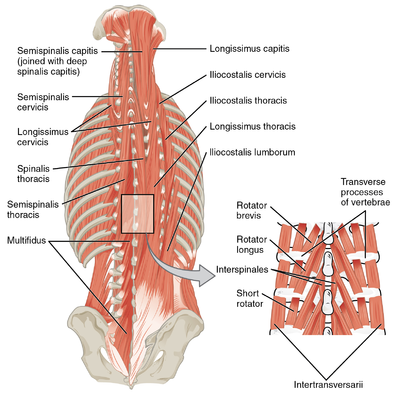
Soft Tissue Injury - Cervical and Lumbar Strain

Lumbar Strain - Physiopedia

Understanding a mass in the paraspinal region: an anatomical approach |  Insights into Imaging | Full Text
Understanding a mass in the paraspinal region: an anatomical approach | Insights into Imaging | Full Text
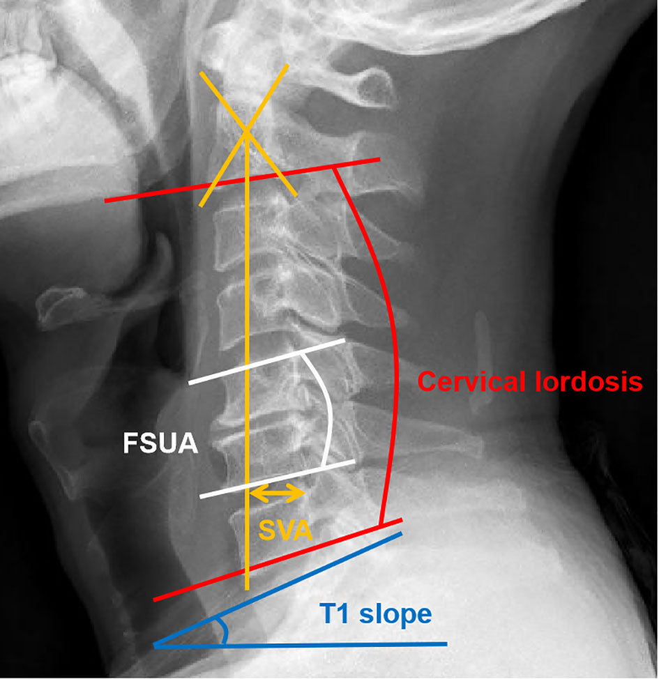
Frontiers | The fatty infiltration into cervical paraspinal muscle as a  predictor of postoperative outcomes: A controlled study based on hybrid  surgery
Frontiers | The fatty infiltration into cervical paraspinal muscle as a predictor of postoperative outcomes: A controlled study based on hybrid surgery

Normal Thickness and Appearance of the Prevertebral Soft Tissues on  Multidetector CT | American Journal of Neuroradiology
Normal Thickness and Appearance of the Prevertebral Soft Tissues on Multidetector CT | American Journal of Neuroradiology

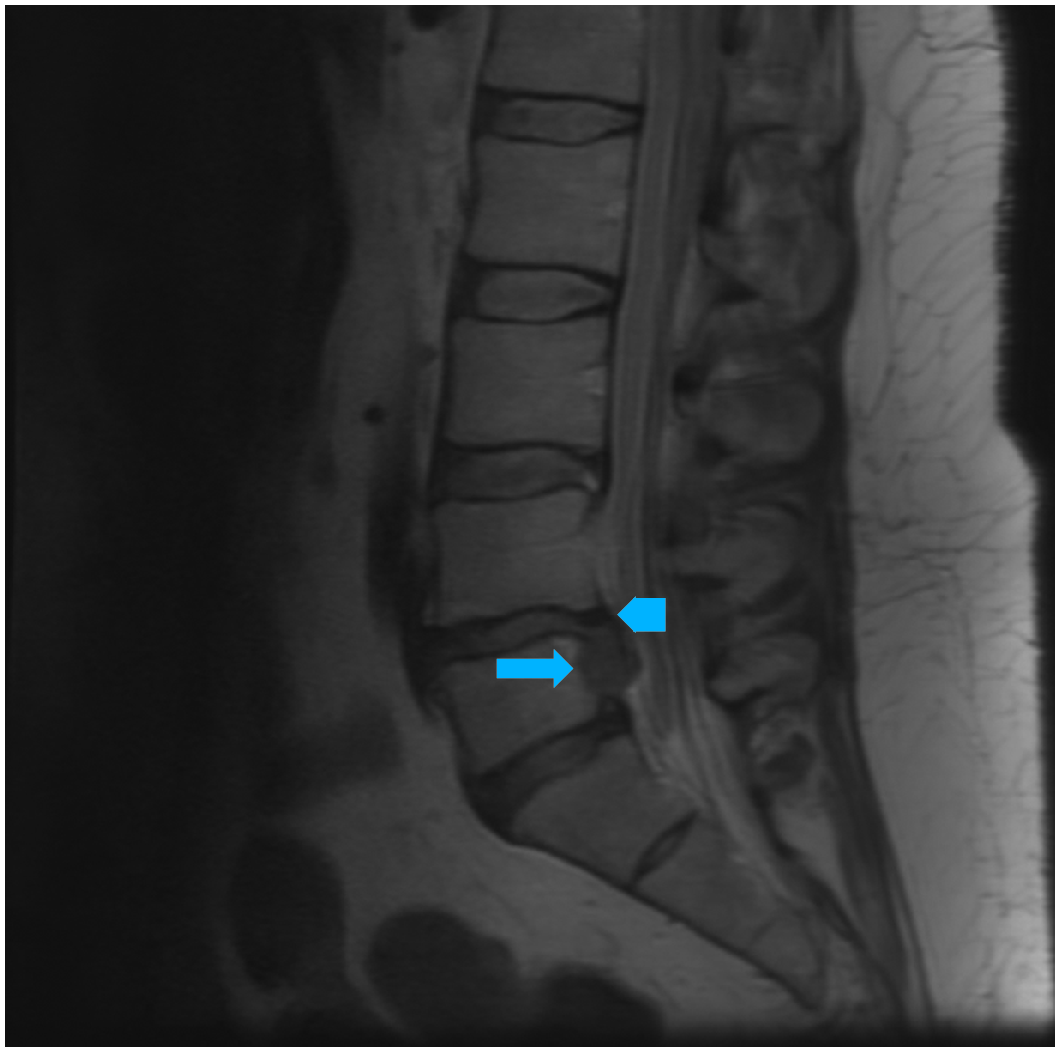
Paraspinal soft tissue edema ratio: An accurate marker for early lumbar  spine spondylodiscitis on an unenhanced MRI - ScienceDirect
Paraspinal soft tissue edema ratio: An accurate marker for early lumbar spine spondylodiscitis on an unenhanced MRI - ScienceDirect

Understanding a mass in the paraspinal region: an anatomical approach |  Insights into Imaging | Full Text
Understanding a mass in the paraspinal region: an anatomical approach | Insights into Imaging | Full Text

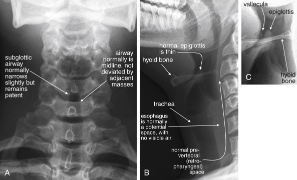
Normal Thickness and Appearance of the Prevertebral Soft Tissues on  Multidetector CT | American Journal of Neuroradiology
Normal Thickness and Appearance of the Prevertebral Soft Tissues on Multidetector CT | American Journal of Neuroradiology